Dorsolateral Prefrontal and Anterior Cingulate Cortex Volumetric Abnormalities in Adults with Attention-Deficit/Hyperactivity Disorder Identified by Magnetic Resonance Imaging
What differences in brain structure can be seen in adults with ADHD?
Larry J. Seidman, Eve M. Valera, Nikos Makris, Michael C. Monuteaux, Denise L. Boriel, Kalika Kelkar, David N. Kennedy, Verne S. Caviness, George Bush, Meg Aleardi, Stephen V. Faraone, and Joseph Biederman


Objectives
What differences can be seen in the brains of people with ADHD?
Previous studies have shown differences in the volume of white matter and gray matter in children with ADHD.
However, before this study, there was not as much research on the brain in adult ADHD.
Researchers in this study wanted to use MRI, an imaging technique that lets scientists look inside the brain, to find if there were any consistent differences in adults with ADHD compared to those without ADHD.
Gray and white matter volume deficits have been reported in a number of studies of children with attention-deficit/ hyperactivity disorder (ADHD); however, there is a paucity of structural magnetic resonance imaging (MRI) studies of adults with ADHD. This structural MRI study used an a p...
Methods
Researchers recruited 24 adults with ADHD and 18 adults without ADHD for the study.
They scanned each of these participants with an MRI and compared the results while accounting for differences in age, sex, and overall brain volume.
Twenty-four adults with DSM-IV ADHD and 18 healthy controls comparable on age, socioeconomic status, sex, handedness, education, IQ, and achievement test performance had an MRI on a 1.5T Siemens scanner. Cortical and sub-cortical gray and white matter were segmented. Image parcellation d...
Results
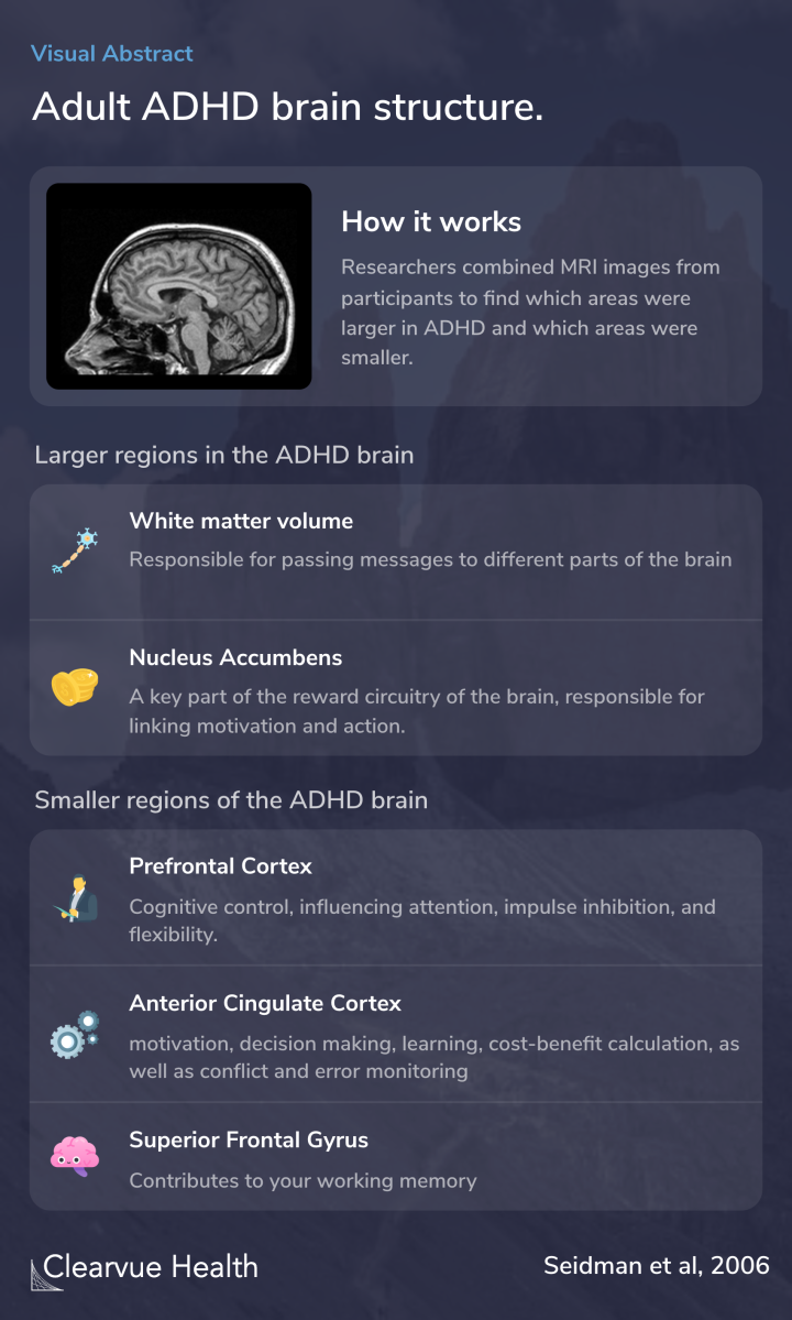
Overall, those with ADHD had more white matter and less gray matter in their brains.
White matter is the part of the brain responsible for passing messages to other parts of the brain.
Those with ADHD also had more volume in their nucleus accumbens, a key part of the brain’s reward system. This part of the brain helps you feel good when you do things that you want to do. Some would say that it links reward with action. However, it can also play a role in addiction.
They were also parts of the brain with less volume in adult ADHD. The prefrontal cortex, responsible for higher-level control in your brain, generally appears smaller in those with ADHD. This is consistent with the fact that those with ADHD have trouble with attention and inhibiting their impulses.
There were also significant differences seen in the superior frontal gyrus and the anterior cingulate cortex. Your superior frontal gyrus is responsible for your working memory. The anterior cingulate cortex is responsible for impulse control and attention.
Relative to controls, ADHD adults had significantly smaller overall cortical gray matter, prefrontal and ACC volumes.
Conclusions
The brain regions found to be different in those with ADHD correspond to the key symptoms of ADHD.
Impulse control, working memory, and attention are all defining features of ADHD, as seen in the diagnostic criteria below.
These data offer further evidence that adult ADHD is a real disorder. The symptoms of adult ADHD have a biological basis that can be seen on MRI. There are physical changes in the brain that correspond to psychological symptoms of ADHD.
Adult ADHD has not always been widely recognized. This study, along with several others, has helped adult ADHD gain recognition as a valid psychiatric condition.
Adults with ADHD have volume differences in brain regions in areas involved in attention and executive control. These data, largely consistent with studies of children, support the idea that adults with ADHD have a valid disorder with persistent biological features.